Radionuclide imaging in mastology
1. Radionuclide diagnostic imaging for breast cancer
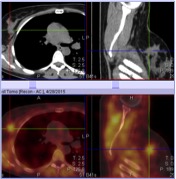
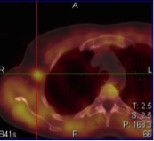
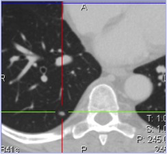
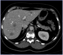
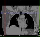
Types of tests: planar mammoscintigraphy, SPECT/CT of the breast, SPECT/CT of the breast and the abdomen with intravenous contrast enhancement, using an iodine-containing contrast agent
Aim: to diagnose and stage breast cancer; to diagnose recurrent cancers; to assess the efficacy of breast cancer treatment
Indications:
- to additionally assess the condition of the liver and lungs under intravenous contrast enhancement with an iodine-containing contrast agent.
- to determine the efficacy of chemotherapy and radiation therapy in patients with breast cancer
- To diagnose recurrences of the disease after surgical interventions
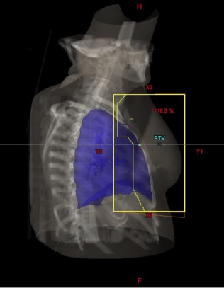
- To prepare a patient for radiation therapy and plan radiation therapy
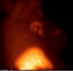
Imaging agent: Tc99m-MIBI (Technetium (99mTc) sestamibi)
Contrast agent for CT: non-ionic iodine containing contrast medium
Principles: Concentration of the imaging agent in the tumor cells (mitochondria) by passive diffusion. The test is performed 5-15 minutes after an intravenous injection of the imaging agent.
Nuances: The imaging agent is injected in a vein on one of the patient’s feet.
Preparation: No special preparation is necessary.
SPECT/CT allows more accurate detection of a tumor in the breast and metastases in the lymph glands of the armpit. SPECT/CT with intravenous contrasting using an iodine-containing contrast agent allows to make comprehensive assessment of the condition of the breast, lymph nodes and the organs of the chest and abdomen.
2. Radionuclide diagnostic imaging of sentinel lymph nodes
Types of tests: mammoscintigraphy of sentinel lymph nodes, SPECT/CT of sentinel lymph nodes
Aim: to visualize sentinel lymph nodes and lymphatic drainage pathways with subsequent operative histodiagnosis of micrometastases
Indications:
- To diagnose metastases and lymphatic drainage pathways in patients with melanoma, tongue cancer, endometrial cancer and cervical cancer.
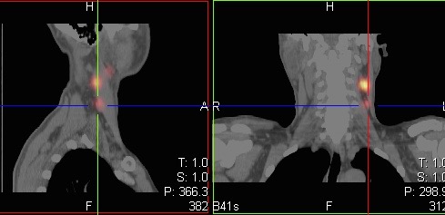
Imaging agent: Technetium-99m sulfur colloid. When injected into a tumor, the particles of the imaging agent migrate along lymphatic drainage pathways into sentinel lymph nodes.
Principle: The imaging agent is injected into the tumor or near it in cases of melanoma. The area of regional lymphatic drainage pathways can be visualized 5-30 minutes after an injection. During surgery, the located sentinel lymph nodes are identified using a special detecting device, removed and sent to a rapid histopathological examination.
Preparation: No special preparation is necessary.
Nuances: The test is to be performed only on condition that the clinical data and other imaging techniques (UT, MRI) have not revealed overt metastases of the regional lymph nodes.
SPECT/CT allows precise location of sentinel lymph nodes.